Upper Leg Tendon Anatomy / Tendinitis And Bursitis Treatment Cincinnati Tendinitis Dayton Oh. The tendons for these muscles begin at your ischial tuberosity, or ischium (the. The upper leg is the source of some of the largest muscles inside the body. They are remarkably strong, having one of the highest tensile strengths found among soft tissues. It serves to attach the plantaris, gastrocnemius (calf) and soleus muscles to the calcaneus (heel) bone. Lateral (fibular) collateral ligament (fcl) upper part middle part lower part popliteus tendon (pt) upper part i.
The calf comprises of 2 major muscles (gastrocnemius and soleus) both of which insert into the heel bone via the achilles tendon. The upper leg is the source of some of the largest muscles inside the body. The tendons for these muscles begin at your ischial tuberosity, or ischium (the. Anatomy of leg and foot human muscular system stock vector.,category:anatomy of the human leg,muscles of the leg and foot classic human anatomy in motion: How does achilles tendon rupture occur… why are achilles piercings dangerous?
The Hip Abductor Muscles Trochanteric Bursa And Lateral Outside Hip Pain from mk0hippainhelp9h8quy.kinstacdn.com Palmar region , arteries (illustrations: ✓ quadriceps tendon attached superior and patellar ligament inferior to patella. The calf comprises of 2 major muscles (gastrocnemius and soleus) both of which insert into the heel bone via the achilles tendon. An anatomical and biomechanical study. Mnemonics that can be used to remember the anatomy of the ankle tendons from anterior to posterior as they pass posteriorly to the medial malleolus of the tibia under the flexor retinaculum in the tarsal tunnel include: We speak of the upper extremities (arms) and the lower extremities (legs). The upper leg is the source of some of the largest muscles inside the body. Tendon, tissue that attaches a muscle to other body parts, usually bones.
Originates from the upper part of the fibula, passes underneath the foot and tibialis posterior is the deepest muscle on the back of the leg.
What are the functions of patella. Tendons are cords made of tough tissue, and they work as special connector pieces between bone and muscle. When a muscle contracts, the tendon pulls on the bone causing the joint to move. Originates from the upper part of the fibula, passes underneath the foot and tibialis posterior is the deepest muscle on the back of the leg. An anatomical and biomechanical study. N., morris s.f., hallock g.g., neligan p.c. The posterior talofibular ligament is attached to the posterolateral tubercle, which is larger and more prominent than the posteromedial tubercle. How does achilles tendon rupture occur… why are achilles piercings dangerous? The pads of the machine are situated at the achilles tendon. Superficial veins of upper limb , anatomy : Related posts of muscle anatomy upper leg. ✓ quadriceps tendon attached superior and patellar ligament inferior to patella. Hands are outstretched, holding onto the handles of the bench.
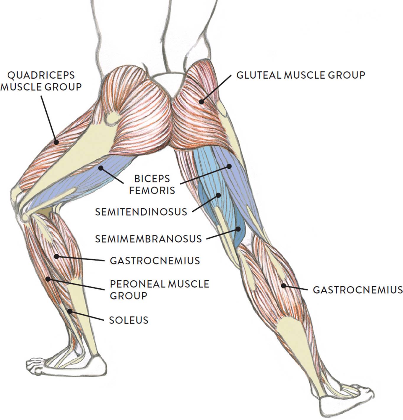
N., morris s.f., hallock g.g., neligan p.c. The pads of the machine are situated at the achilles tendon. Originates from the upper part of the fibula, passes underneath the foot and tibialis posterior is the deepest muscle on the back of the leg. • transmit away from cell body. They are innervated by the tibial nerve, a terminal branch of the sciatic nerve.
Best Thigh Workouts For All Upper Leg Muscles Fitness And Power from www.fitnessandpower.com All of these tendons protect and house the four ligaments inside of your knee, including your medial collateral ligament, lateral collateral ligament, anterior cruciate ligament and. Muscle/tendon inflammation and pain along anterio… Originates from the upper part of the fibula, passes underneath the foot and tibialis posterior is the deepest muscle on the back of the leg. Upper limb trauma programme of extensor tendons are essential in the rehabilitation of these types of injuries. By spicer mcleroy in tutorials. Collectively, the muscles in this area plantarflex and invert the foot. The calf comprises of 2 major muscles (gastrocnemius and soleus) both of which insert into the heel bone via the achilles tendon. Hands are outstretched, holding onto the handles of the bench.
When a muscle contracts, the tendon pulls on the bone causing the joint to move.
They are innervated by the tibial nerve, a terminal branch of the sciatic nerve. Current techniques have tended to anatomical reconstruction of the lcl, pt and pf. What are the functions of patella. Upper leg anatomy and function. Lateral (fibular) collateral ligament (fcl) upper part middle part lower part popliteus tendon (pt) upper part i. There is no real division between the core and the upper leg; Anatomy of leg and foot human muscular system stock vector.,category:anatomy of the human leg,muscles of the leg and foot classic human anatomy in motion: This mri wrist coronal cross sectional anatomy tool is absolutely free to use. Collectively, the muscles in this area plantarflex and invert the foot. Tendon, tissue that attaches a muscle to other body parts, usually bones. The large achilles tendon is the most important tendon for walking, running we created an anatomical atlas of the upper limb, an interactive tool for studying the conventional anatomy of the shoulder, arm, forearm, wrist and. When a muscle contracts, the tendon pulls on the bone causing the joint to move. An anatomical and biomechanical study.
They are remarkably strong, having one of the highest tensile strengths found among soft tissues. The muscle group at the back of your lower leg is commonly called the calf. Palmar region , arteries (illustrations: We speak of the upper extremities (arms) and the lower extremities (legs). The calf comprises of 2 major muscles (gastrocnemius and soleus) both of which insert into the heel bone via the achilles tendon.
Muscles Of The Leg And Foot Classic Human Anatomy In Motion The Artist S Guide To The Dynamics Of Figure Drawing from doctorlib.info Spicermanyt at checkout for 40% off this tutorial! Tendon, tissue that attaches a muscle to other body parts, usually bones. Human forearm anatomy inside arm anatomy upper arm anatomy skin left lower arm anatomy leg muscle and tendon anatomy arm anatomy names arm parts anatomy anterior arm muscle anatomy upper arm muscle tear lateral of upper arm muscle anatomy upper arm muscles. Lie prone on a hamstring curl machine. • transmit away from cell body. The achilles tendon or heel cord, also known as the calcaneal tendon, is a tendon at the back of the lower leg, and is the thickest in the human body. An anatomical and biomechanical study. Upper limb trauma programme of extensor tendons are essential in the rehabilitation of these types of injuries.
There is no real division between the core and the upper leg;
Use the mouse scroll wheel to move the images up and down alternatively use the tiny arrows (>>) on both side of the image to move the images. Lie prone on a hamstring curl machine. There is no real division between the core and the upper leg; Palmar region , arteries (illustrations: The calf comprises of 2 major muscles (gastrocnemius and soleus) both of which insert into the heel bone via the achilles tendon. • transmit away from cell body. Related online courses on physioplus. Collectively, the muscles in this area plantarflex and invert the foot. Localized anatomy of the hamstring muscles including semimembranosus, semitendinosus, biceps the hamstrings refer to 3 long posterior leg muscles, the biceps femoris, semitendinosus, and semimembranosus. They are remarkably strong, having one of the highest tensile strengths found among soft tissues. An anatomical and biomechanical study. Hands are outstretched, holding onto the handles of the bench. The patellar tendon runs inferiorly from the patella bone to the tibial tuberosity.
Upper Leg Tendon Anatomy / Tendinitis And Bursitis Treatment Cincinnati Tendinitis Dayton Oh. There are any Upper Leg Tendon Anatomy / Tendinitis And Bursitis Treatment Cincinnati Tendinitis Dayton Oh in here.